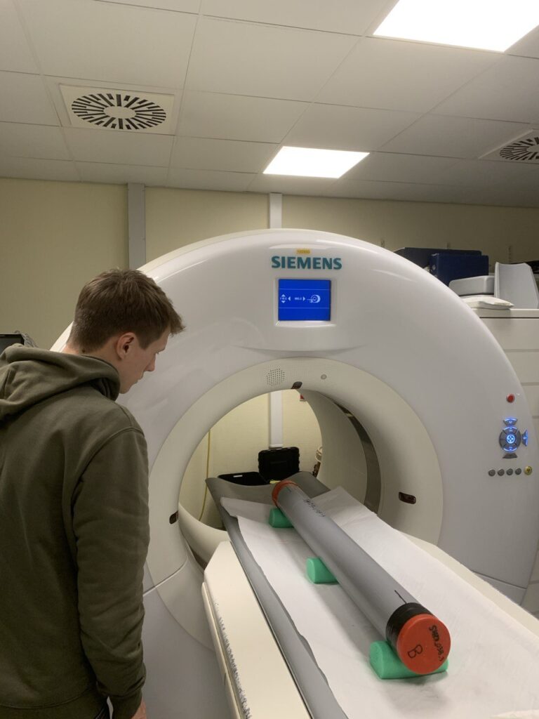
As part of our interdisciplinary approach in the NORSEAT Project, our team recently collaborated with Ghent University Hospital to conduct medical CT scanning on sediment cores. This advanced imaging method allows us to investigate the internal structure, grain distribution, and sediment composition in unprecedented detail—without disturbing the cores themselves.
Why Use a Medical CT Scanner?
Typically used for human diagnostics, medical CT (Computed Tomography) scanning offers ultra-high-resolution, three-dimensional imaging. Applied to sediment cores, this technology helps us:
- Visualize the internal grain structure and layering,
- Detect variations in density and grain size distribution,
- And build 3D models to guide targeted sampling and stratigraphic interpretation.
This level of precision is invaluable for detecting subtle features like tsunami-related deposits or sedimentary disturbances.
Imaging in Action
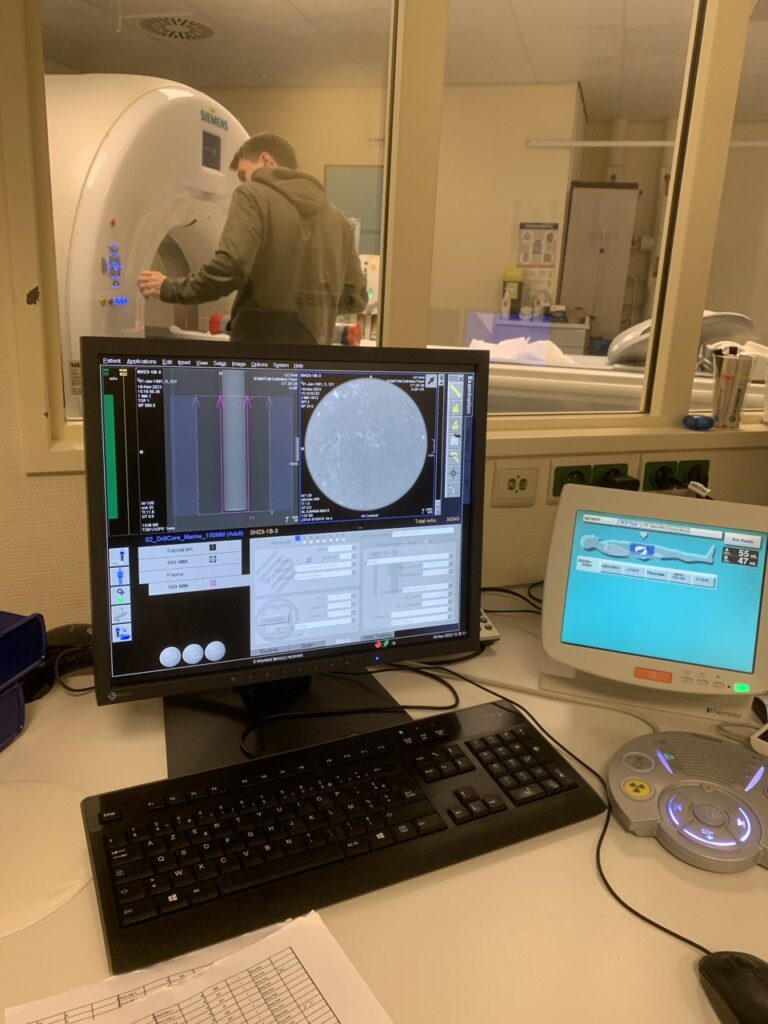
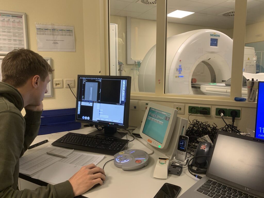
The CT scans were conducted on full-length sediment cores still encased in their plastic liners, ensuring preservation of core integrity. The results produced high-resolution 3D models and cross-sectional images, revealing intricate details of grain composition and internal layering that are often missed by traditional imaging techniques.
Bridging Earth Science and Medical Technology
This collaboration highlights the power of cross-disciplinary innovation, combining geological expertise with cutting-edge medical imaging. The results not only improve our understanding of sediment deposition dynamics in the Shetland offshore zone, but also pave the way for more accurate reconstructions of palaeoenvironmental events, including tsunamis.
Stay tuned for more behind-the-scenes looks into our research methods as the NORSEAT Project continues to push boundaries in Earth science.